Diagram Of The Muscles In The Forearm : Muscle Compartments Of The Forearm Complete Anatomy
ads/wkwkland.txt
Diagram Of The Muscles In The Forearm : Muscle Compartments Of The Forearm Complete Anatomy. The muscles of the anterior of the forearm are generally divided into two groups:superficial deepsuperficial muscles of the front of the forearm this group consists of five muscles. 4, attachment… the muscles of the back forearm. A helpful way to learn anatomy is to move and mimic the actions for the muscles you are learning that week. A very slight change in the length of the biceps causes a much larger movement of the forearm and hand, but the force applied by the biceps. 3d anatomy tutorial on the muscles of the flexor compartment of the forearm.
ads/bitcoin1.txt
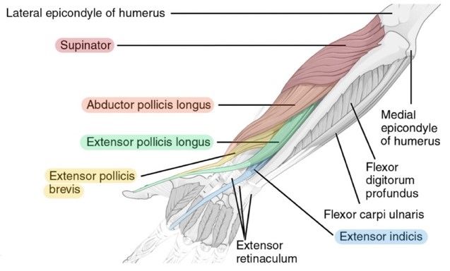
The superficial extensors of the forearm are the brachioradialis, extensor carpi radialis longus, anconeus, extensor carpi radialis brevis, extensor carpi ulnaris, extensor digitorum and extensor digiti minimi. Flexor carpi ulnaris, palmaris longus, flexor carpi radialis, and pronator teres. Remembering the action of each one can be quite difficult. Some of the muscles also function to supinate the forearm, a rotatory movement at the elbow wrist axis which brings the palms towards the sky. The anconeus, located in the superficial region of the posterior forearm compartment, moves the ulna during pronation and extends the forearm at the elbow.
Muscles Of The Forearm Labeled Diagram Poster Zazzle Com from rlv.zcache.com Learn vocabulary, terms and more with flashcards, games and other study tools. In the anterior compartment, they are split into three categories: 2, ulna, 3, biceps muscle; The accompanying muscle diagram reveals the muscles' positions beneath the surface. A deep layer, intermediate layer and superficial layer. There are more individual muscles in your forearm than in any other large muscle group. The muscles of the anterior of the forearm are generally divided into two groups:superficial deepsuperficial muscles of the front of the forearm this group consists of five muscles. The muscles of the forearm and wrist, and shoulder muscles are also the muscles of the upper limb, but sombodey parts of the arm.
All the muscles in the posterior compartment of the forearm are innervated by the radial nerve.
ads/bitcoin2.txt
This is a fusiform muscle that forms the lateral boundary of the cubital fossa and is the most superficial muscle on the radial side of the forearm. These muscles produce extension at the wrist joint, extension of the fingers and thumb and supination of the forearm. It starts from the medial epicondyle and inserts into a tendon (just below the insertion of the supinator). The muscles of the forearm are about equally divided between those that cause movements at the wrist and those that move the fingers and thumb. Each muscle roughly follows the course of digits. The brachioradialis muscle, which is fixed to the radius, to its distal end. Here's an example of a petite woman. Remembering the action of each one can be quite difficult. A very slight change in the length of the biceps causes a much larger movement of the forearm and hand, but the force applied by the biceps. The antibrachial or forearm muscles may be divided into a volar and a dorsal group. 2, ulna, 3, biceps muscle; Diagram the movements of the humerus muscles that act on the forearm. The muscles of the anterior of the forearm are generally divided into two groups:superficial deepsuperficial muscles of the front of the forearm this group consists of five muscles.
There are two parts to this tutorial, this is the first part on the anterior. A deep layer, intermediate layer and superficial layer. There are many muscles in the forearm. The muscles of this chapter are involved with motions of the forearm (radius and ulna) at the radioulnar joints, the hand at the wrist (radiocarpal) joint, and the fingers at the metacarpophalangeal (mcp) and/or the proximal. Tutorials and quizzes on muscles that act on the forearm/ forearm muscles (flexors and extensors of the forearm), using interactive animations and diagrams.
11 Muscles Of The Forearm Simplemed Learning Medicine Simplified from simplemed.co.uk Tutorials and quizzes on muscles that act on the forearm/ forearm muscles (flexors and extensors of the forearm), using interactive animations and diagrams. Learn vocabulary, terms and more with flashcards, games and other study tools. The anterior forearm muscles are divided into 3 muscular layers; The flexor carpi ulnaris lies along the ulnar side of the forearm. The forearm is a mass of some 20 different muscles. Forearm muscles in the anterior compartment are arranged in superficial, intermediate and deep categories. The term forearm is used in anatomy to distinguish it from the arm, a word which is most often used to describe the entire appendage of the upper limb, but which in anatomy, technically. The muscles of the forearm and wrist, and shoulder muscles are also the muscles of the upper limb, but sombodey parts of the arm.
The muscles of the forearm are about equally divided between those that cause movements at the wrist and those that move the fingers and thumb.
ads/bitcoin2.txt
2, ulna, 3, biceps muscle; By simply having the forearm strength to hold greater weight for more time, you can help extend your shoulder, bicep the muscles of the forearm are predominantly slow twitch. .diagram | forearm muscles 13. The superficial extensors of the forearm are the brachioradialis, extensor carpi radialis longus, anconeus, extensor carpi radialis brevis, extensor carpi ulnaris, extensor digitorum and extensor digiti minimi. The anterior forearm muscles are divided into 3 muscular layers; It leads to flexion of the forearm and helps the brush to a position intermediate between. Your arm muscles allow you to perform hundreds of everyday movements, from making a fist to bending your thumb. I've just switched over to a diagram to show you this muscle. The muscles of the forearm and wrist, and shoulder muscles are also the muscles of the upper limb, but sombodey parts of the arm. Look at the picture of the muscle, find it on your body, and picture how it is contracting as it produces its associated movement or movements. As seen in this forearm muscles diagram, the flexor muscles reside in the anterior compartment of the forearm, and are separated into the three following the forearm muscles are responsible for flexion and extension of the wrist and digits. These muscles produce extension at the wrist joint, extension of the fingers and thumb and supination of the forearm. Learning their anatomy will help you design awesomely dynamic arms.
Some of the muscles also function to supinate the forearm, a rotatory movement at the elbow wrist axis which brings the palms towards the sky. Here, we will discuss the anterior compartment of the forearm in the setting of their a neat little trick to learn the superficial muscles of the forearm is to use your fingers as the guide. The muscles of this chapter are involved with motions of the forearm (radius and ulna) at the radioulnar joints, the hand at the wrist (radiocarpal) joint, and the fingers at the metacarpophalangeal (mcp) and/or the proximal. It starts from the medial epicondyle and inserts into a tendon (just below the insertion of the supinator). I've just switched over to a diagram to show you this muscle.
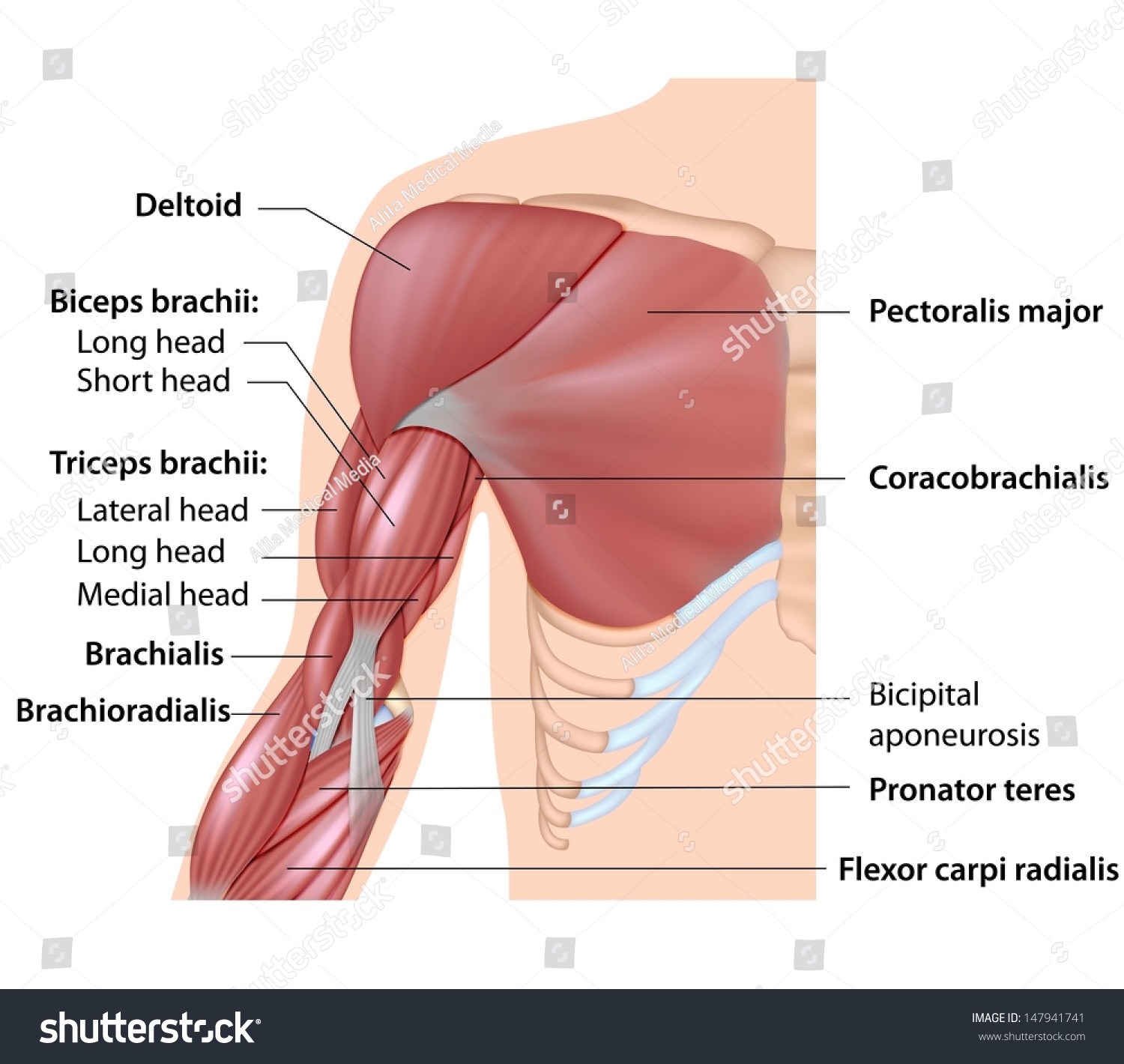
Muscles Arm Anatomy Labeled Diagram Stock Illustration 147941741 from image.shutterstock.com Flexor carpi ulnaris, palmaris longus, flexor carpi radialis, and pronator teres. The flexor carpi ulnaris lies along the ulnar side of the forearm. Learning their anatomy will help you design awesomely dynamic arms. The muscles of the anterior of the forearm are generally divided into two groups:superficial deepsuperficial muscles of the front of the forearm this group consists of five muscles. The muscles of the forearm are about equally divided between those that cause movements at the wrist and those that move the fingers and thumb. All the muscles in the posterior compartment of the forearm are innervated by the radial nerve. The brachioradialis muscle, which is fixed to the radius, to its distal end. Each muscle roughly follows the course of digits.
Learning their anatomy will help you design awesomely dynamic arms.
ads/bitcoin2.txt
A deep layer, intermediate layer and superficial layer. Flexor carpi ulnaris, palmaris longus, flexor carpi radialis, and pronator teres. Try labeling diagrams and worksheets as additional learning aids. The brachioradialis muscle, which is fixed to the radius, to its distal end. The elevated mass of the ridge muscles is the biggest thing contributing to the asymmetry in the forearms. Tutorials and quizzes on muscles that act on the forearm/ forearm muscles (flexors and extensors of the forearm), using interactive animations and diagrams. It arises by two heads, humeral and ulnar, connected by a tendinous arch, beneath which the ulnar nerve and posterior ulnar recurrent artery pass. One of the famous application are prosthetic and. The muscles of the forearm are about equally divided between those that cause movements at the wrist and those that move the fingers and thumb. 4, attachment… the muscles of the back forearm. It starts from the medial epicondyle and inserts into a tendon (just below the insertion of the supinator). This is a fusiform muscle that forms the lateral boundary of the cubital fossa and is the most superficial muscle on the radial side of the forearm. Here's an example of a petite woman.
ads/bitcoin3.txt
ads/bitcoin4.txt
ads/bitcoin5.txt
ads/wkwkland.txt
0 Response to "Diagram Of The Muscles In The Forearm : Muscle Compartments Of The Forearm Complete Anatomy"


0 Response to "Diagram Of The Muscles In The Forearm : Muscle Compartments Of The Forearm Complete Anatomy"
Posting Komentar